Author: Dr. Ricardo Schestatsk
59-year-old male patient.
Chief complaint: Patient had a longitudinal fracture in element 15, with pain, inflammatory signs and infection.
INITIAL EVALUATION
After the initial examination, anamnesis and tomographic examination, the fracture was detected at the root level, as well as inadequate restoration in the distal part of element 14.
TREATMENT PERFORMED
Prior to surgery, an intraoral scan was performed in order to fabricate a temporary crown. Extraction was planned, with as little trauma as possible, using periotomes in order not to traumatize bone and gingival tissue, especially the interproximal papillae. After washing and curettage of the alveolus, an Arcsys 4.3 x 11 mm frictional implant was installed, leaving 3 mm below the buccal gingival margin.
Afterwards, the printed crown was joined to the multifunctional transfer which was previously worn with Opallis Flow composite, in shade A, Ambar Universal APS adhesive and finished with composite polishing spirals. In 60 days, the region was completely healed and the gingival tissue was in excellent condition to make the zirconia crown. The transfer was made from a Scanbody to a mini abutment, and the crown was made and delivered ready in the following appointment.
STEP BY STEP
1. Scanning and design of printed provisional crown;
2. Initial situation;
3. Exodontia, implant, placement of a 2.5 mm mini abutment;
4. Grafting and provisional placement;
5. After dentistry of the neighboring tooth, scanning with Scanbody;
6. Placement of zirconia crown without the need to test.

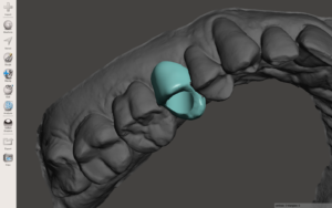
1 e 2 | Scanning and provisional design
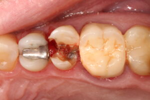
3 | Fractured tooth
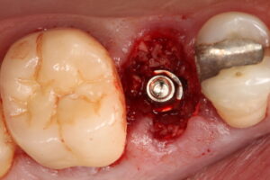
4 | Arcsys implant with excellent stability, grafting and mini abutment
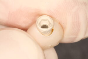
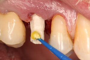
5 e 6 | Capture printed provisional with Opallis Flow composite
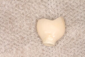
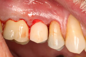
7 e 8 | Provisional finalized and immediate post-surgical
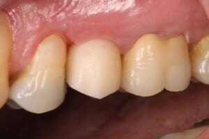
9 | 7 days post surgery
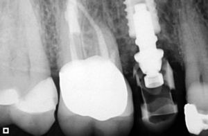
10 | Control x-Ray in 60 days
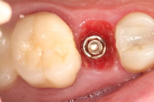
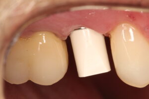
11 e 12 | Tissue stability after 60 days and scanbody in position

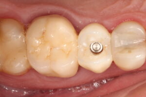
13 e 14 | Final result, after dentistry in element 14
