Authors: Prof. Dr. Everton Salante e Prof. Dr. Leonardo Piazzetta Pellissari
FEMALE PATIENT, 40 YEARS OLD: Changes in element 26 even after endodontic retreatment.
INITIAL EVALUATION
The patient presented a recurrence of periapical lesion after the second endodontic retreatment in tooth 26. The clinical and radiographic evaluation allowed the finding of root resorption with an indication of extraction.
TREATMENT PERFORMED
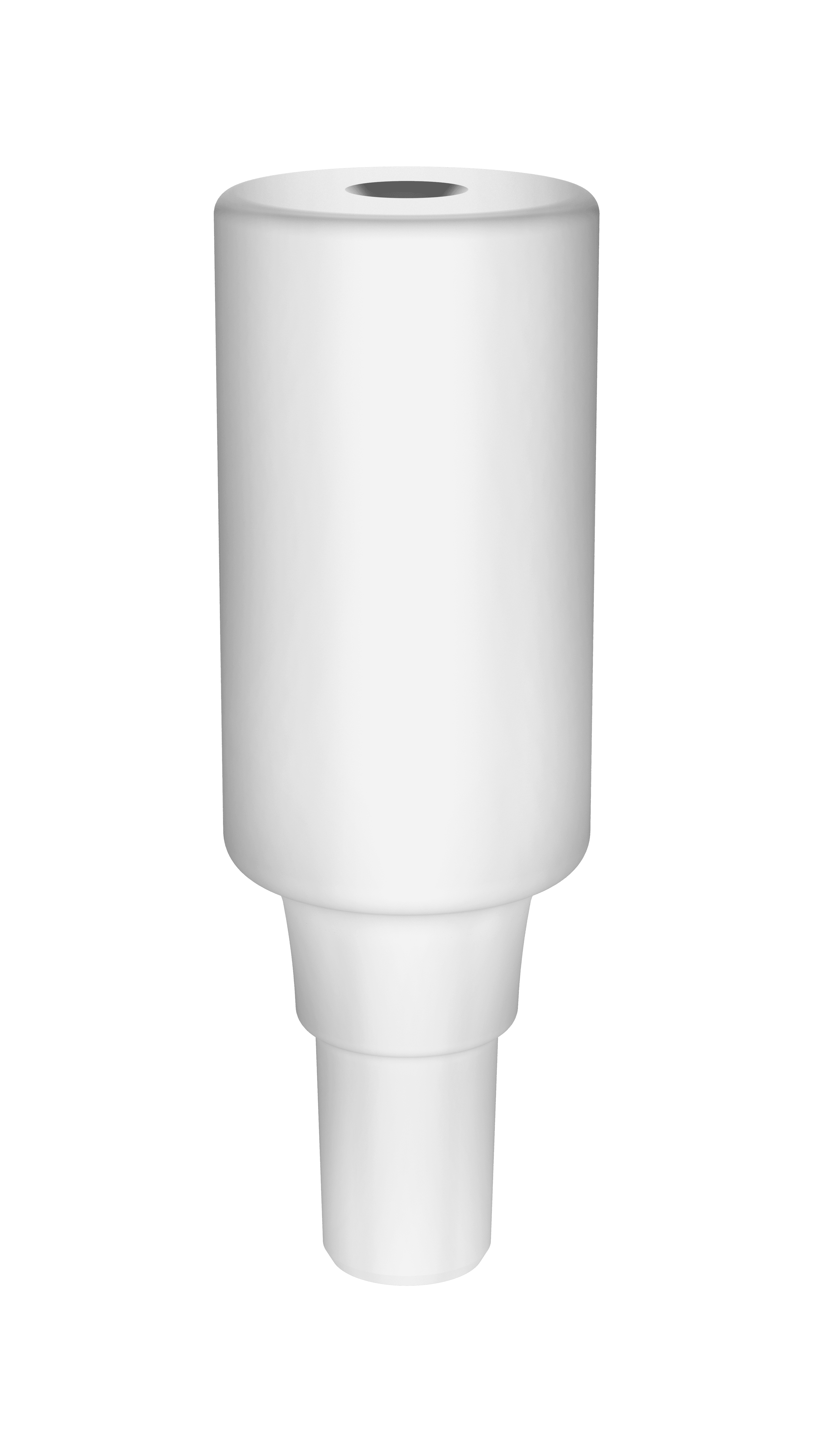
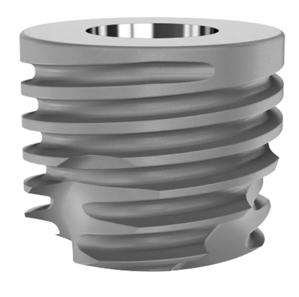
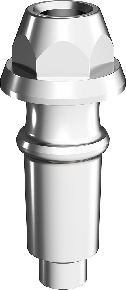
It was proposed to replace the compromised dental element with an immediate implant with immediate loading and custom healing abutment. Thus, a minimally traumatic extraction was performed, preserving the gingival architecture of the region, a 5×5 mm Short Arcsys implant, 2 mm infraosseous, where it reached 32 N.cm of primary stability. Thus, the custom healing abutment was made using a multifunctional high-profile healing abutment with 4 mm of custom diameter, photoactivated pattern resin was added, completely sealing the embouchure of the alveolus. Before the installation of the healing abutment, the implant cover was inserted to prevent biomaterial residues from occupying the internal space of the implant.
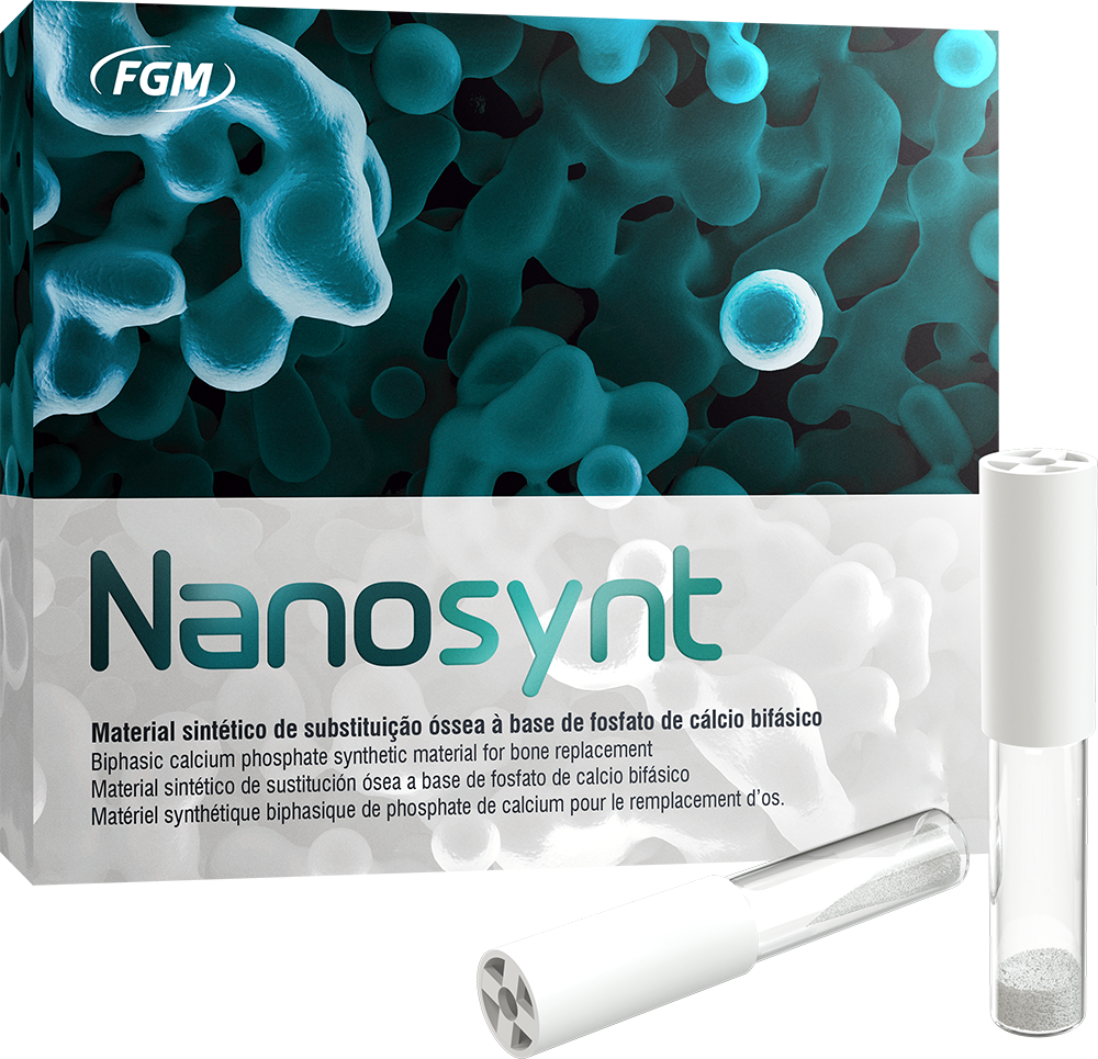
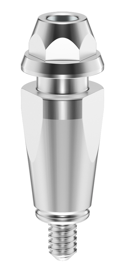
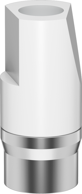
Then the gap was filled with Nanosynt. Then, the implant cover was removed, and the custom healing abutment was immediately and carefully installed with only a digital pressure. As an advantage of this procedure, it can be noted the obliteration of the alveolar embouchure with concomitan tissue conditioning, starting the customization of the emergency profile. After 90 days postoperatively, the healing abutment was removed, and the abutment for screw-retained restoration was installed with 2.5 mm of transmucosal. The peri-implant tissues already had a satisfactory emergency profile to start intraoral scanning. The scan body for the abutment for screw-retained restoration was installed on it, and the .stl files were sent to the prosthetic laboratory, which made a zirconia crown on a metallic link to offer greater resistance and avoid the occurrence of fracture of the crown installed directly over the prosthetic component.
The crown was cemented to the link using Allcem Core cement. Finishing was performed on the interface region of the link with the prosthetic crown, and the assembly was installed on the abutment for screw-retained restoration with a torque of 10 N.cm. On the screw, the Teflon tape and Vittra APS Unique resin were inserted.
STEP BY STEP
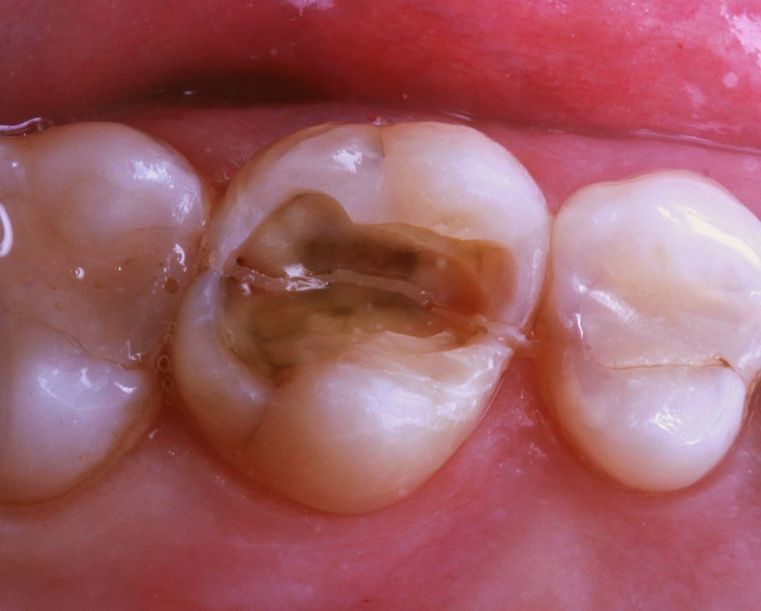
01 – Initial situation
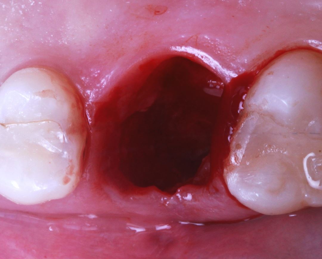
02 – Alveolus after extraction.
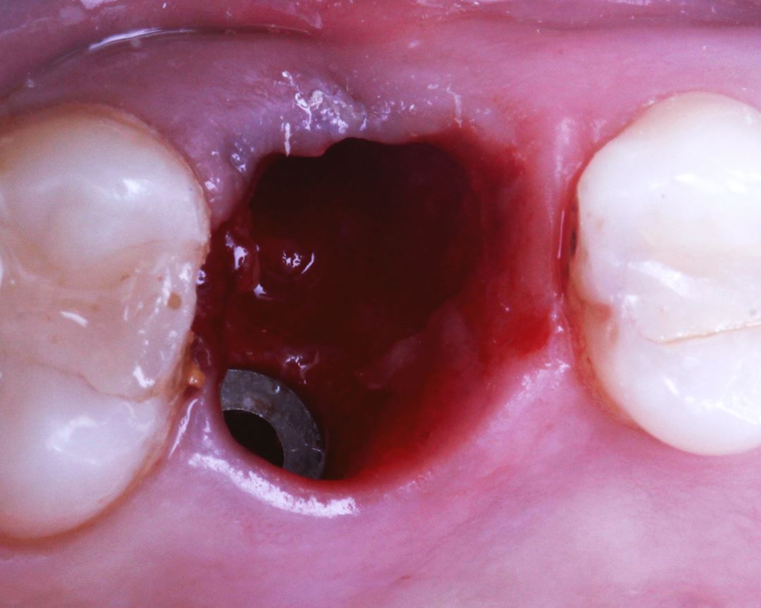
03 – Short implant installation
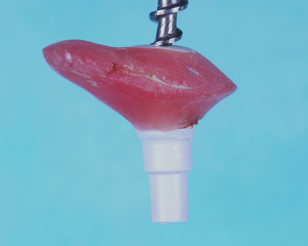
04 – Custom multifunctional healing abutment.
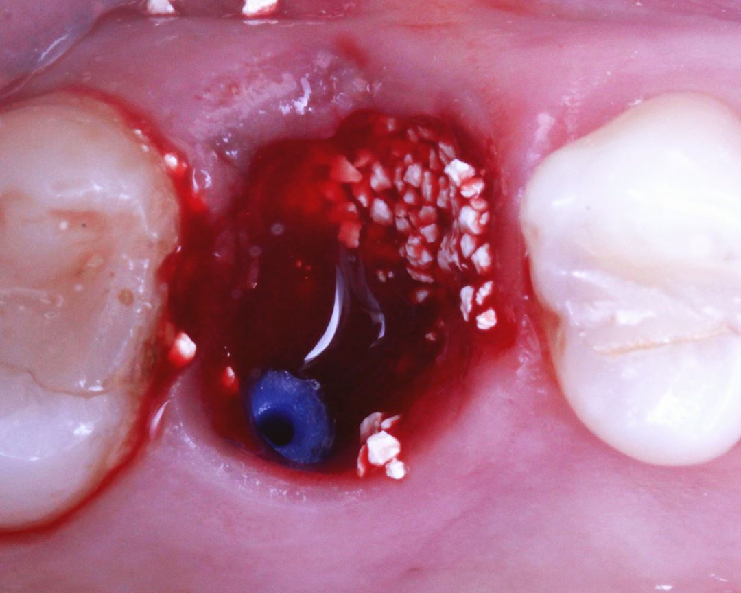
05 – Filling the gap with Nanosynt.
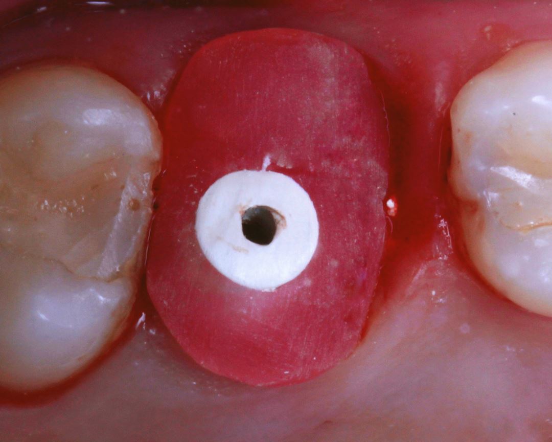
06 – Custom healing abutment installation with Vittra APS.
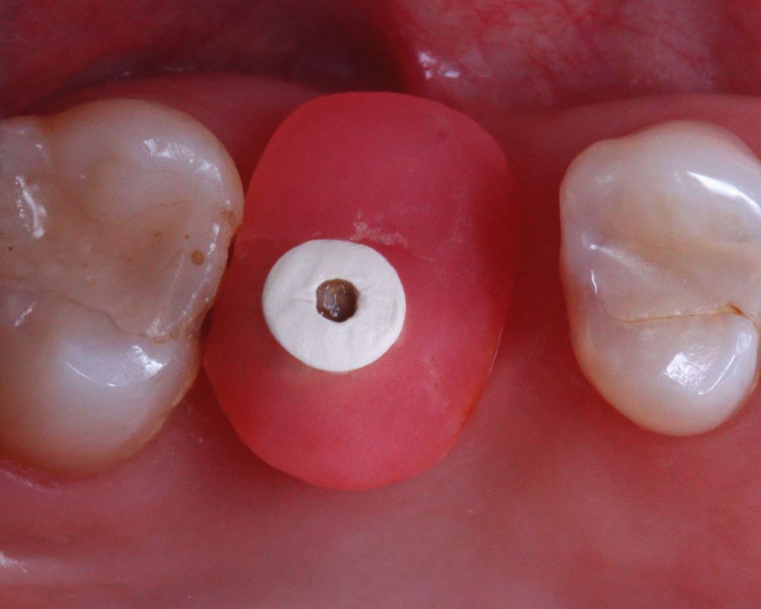
07 – 7-day postoperative period.
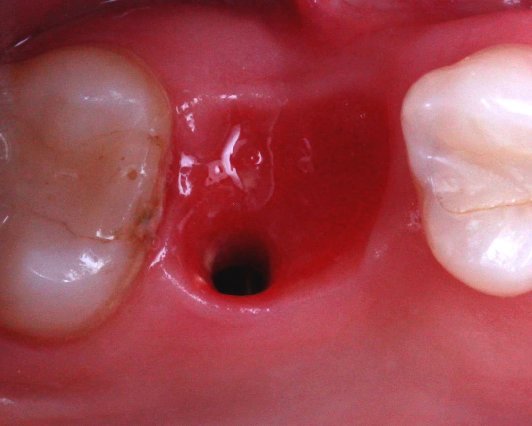
08 – Gingival aspect after three months.
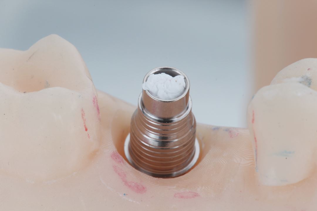
09 – Metallic link on the prosthetic component.
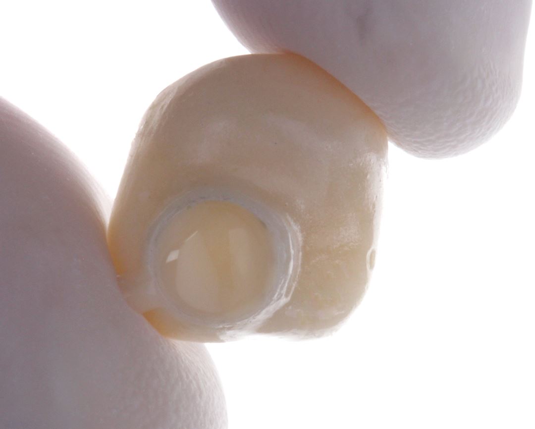
10 – Cement on the inside of the crown.
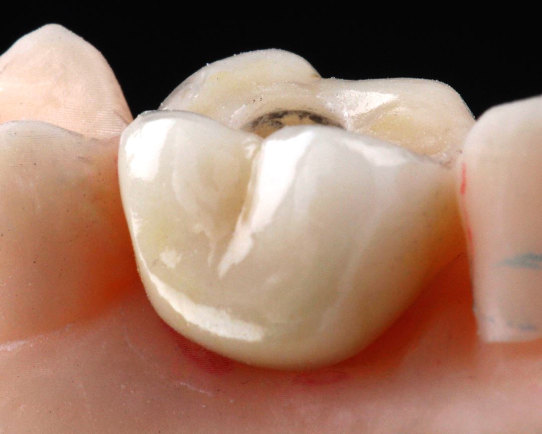
11 – Crown in position on printed model.
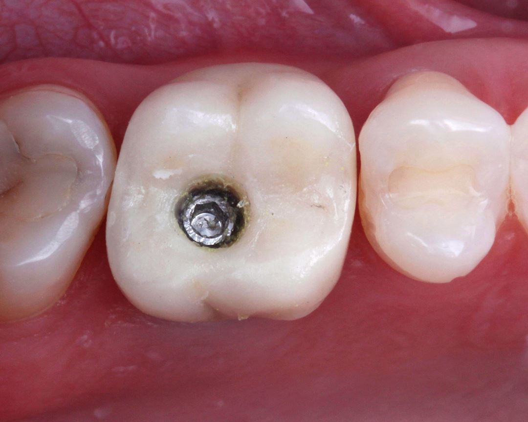
12 – Final situation before applying the resin.
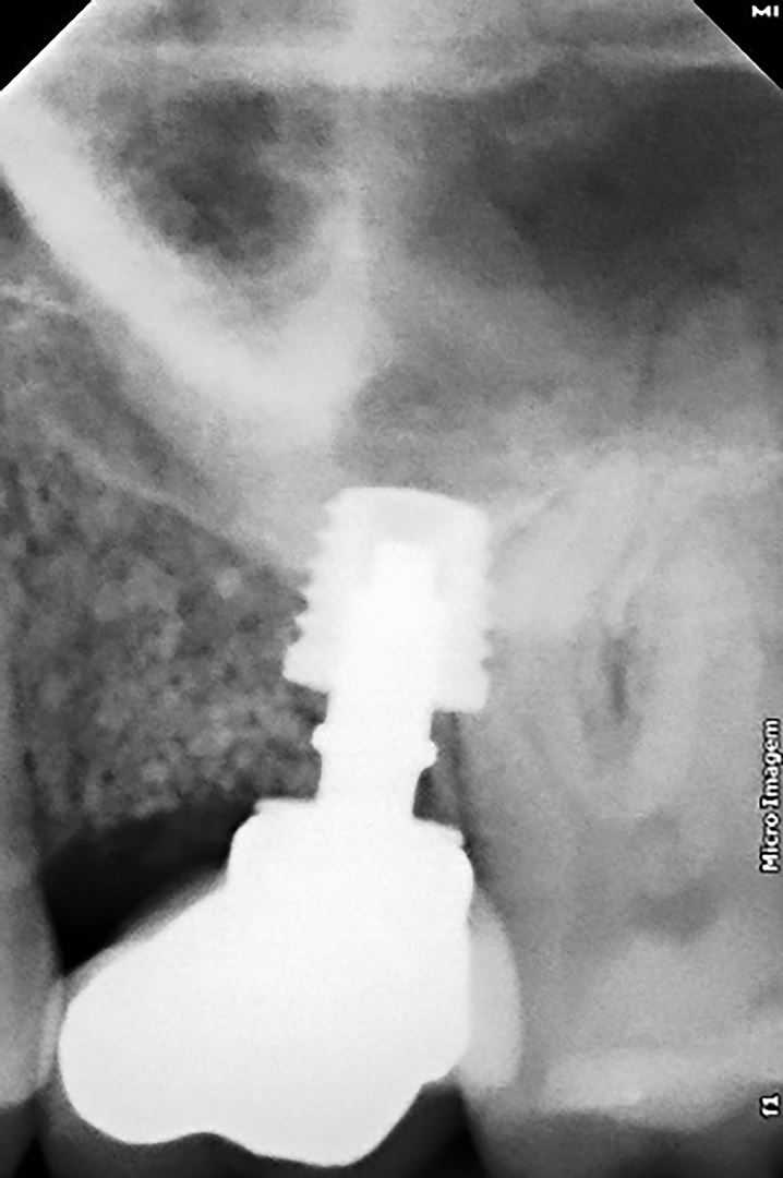
13 – Final radiographic aspect.

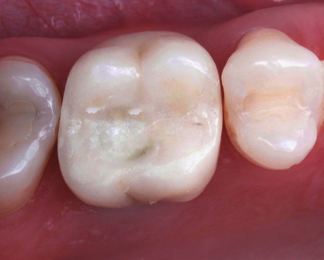
14 e 15 – Initial and final aspect
PRODUCTS USED
