Authors: Dr. Jessica Grassi, Dr. Eduarda Blasi Magini and Dr. Bernardo Born Passoni
32-year-old male patient.
CHIEF COMPLAINT
Pain in the right upper central incisor after a few years of occlusal trauma.
INTRODUCTION
It is widely known that the esthetics of the smile has a direct impact on the patient’s self-esteem. Thus, installation of the immediate unitary implant with immediate provisioning in patients with compromised teeth has become routine in implantology (Balderrama et al, 2021), being an excellent alternative when it is necessary to unite esthetics, predictability, and patient-practitioner satisfaction. Indications for minimally traumatic extraction and immediate implant installation include: teeth with irreversible endodontic treatment failures, teeth with advanced periodontal disease, root fractures, and advanced caries below the gingival margin (Freitas et al, 2019). Dental implants performed immediately after exodontia have a high success rate and allow the rehabilitation of the patient with a smaller amount of interventions for the completion of the clinical case (Medeiros et al. 2020).
Osseointegrated implants can be surgically installed in several temporal phases after tooth extraction. The advantages of immediate implants include: significant reduction in total treatment time; use of immediate fixed prosthesis after implant fixation; reduction of the risk of trauma to implants by the temporary prosthesis; elimination of the transient removable prosthesis; psychological and esthetic benefits for patients; and better bone healing and modulation of the anatomy of adjacent soft tissues (Silva et al. 2021). This technique consists of the removal of a dental element and immediate installation of an implant in the still fresh alveolus. The recommended surgical technique does not require mucoperiosteal incisions or detachments, maintaining the vascularization of the vestibular bone, minimizing its resorption and preserving the interdental papillae (Carneiro et al. 2014).
For the installation of the immediate implants, it is important that the professional evaluate the quantity and quality of the bone, patient occlusion, presence or not of deleterious habits, surgical technique, and general health of the patient. In addition to the installation of the implant, another very important phase for functional and esthetic rehabilitation of the implant is the making of temporary prosthesis, because, with immediate provisioning of implants, gingival conditioning can be performed to receive the definitive crown, obtaining a natural shape of the emergence profile, in addition to meeting the esthetic demands of the patient shortly after surgery (Balderrama et al. 2021).
Tissue conditioning and the correct evaluation of the patient’s gingival phenotype is important during treatment planning. A good soft tissue condition is necessary for the longevity of dental implant treatments and is even more important for implant therapies when considering esthetic factors. This is because the peri-implant soft tissues are similar to the protective periodontium, which provides an important barrier against bacterial aggression to bone tissue. (Nagai et al, 2021).
CASE REPORT
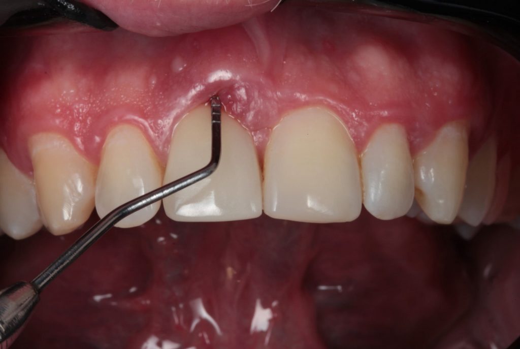
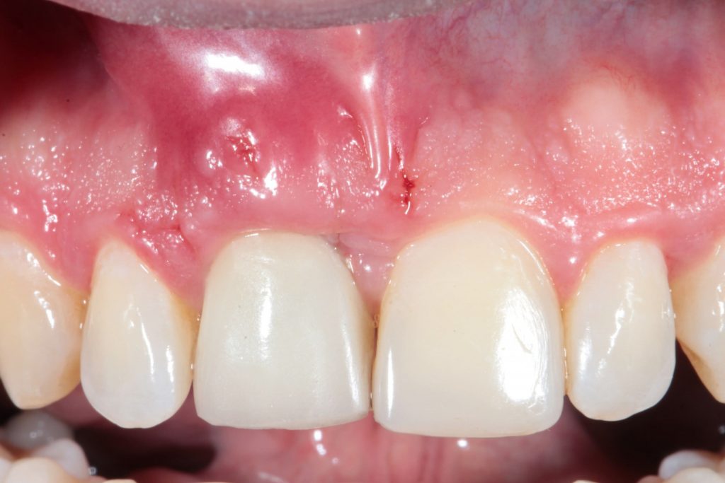
Male patient, 32 years old, leucoderma, without health complications or basic diseases or allergies,came to the private clinic reporting pain in the right upper central incisor after a few years of occlusal trauma. Upon clinical examination, marginal gum hyperemia and vestibular probing depth of 12 mm were observed (Figure 1).
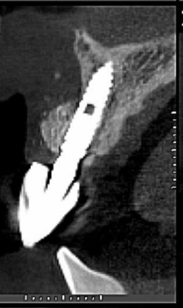
During the anamnesis, he reported that he could not do without a missing tooth in that region, due to esthetic and functional impairment in the area. For the planning of the case, a Cone Beam computed tomography of the region of the dental element 11 was requested. When evaluating the exam image, external resorption was visualized, and the extraction of the dental element with immediate installation of the implant was indicated.
Under local anesthesia (Articaine 4% with epinephrine 1:100,000) via infiltrative terminal in the vestibular region and infiltrative anesthesia of the nasopalatin, intrasucular incision, and syndesmotomy without papilla detachment, extraction was performed in a minimally traumatic way with the use of a periotome. After the extraction, single milling was performed with 2.4 mm drill for 3.3 x 11 Arcsys FGM Morse Taper implant installation. The implant was positioned through the Palatal approach and at a distance of 2 mm from the vestibular roof and 5 mm from the gingival margin. This coronal apex positioning is essential for the correct formatting of biological distances, as the morse taper implant must be 2 mm infraosseous, added to the 3 mm for the formation of biological distances (sulcus epithelium, junctional epithelium, and connective adaptation).
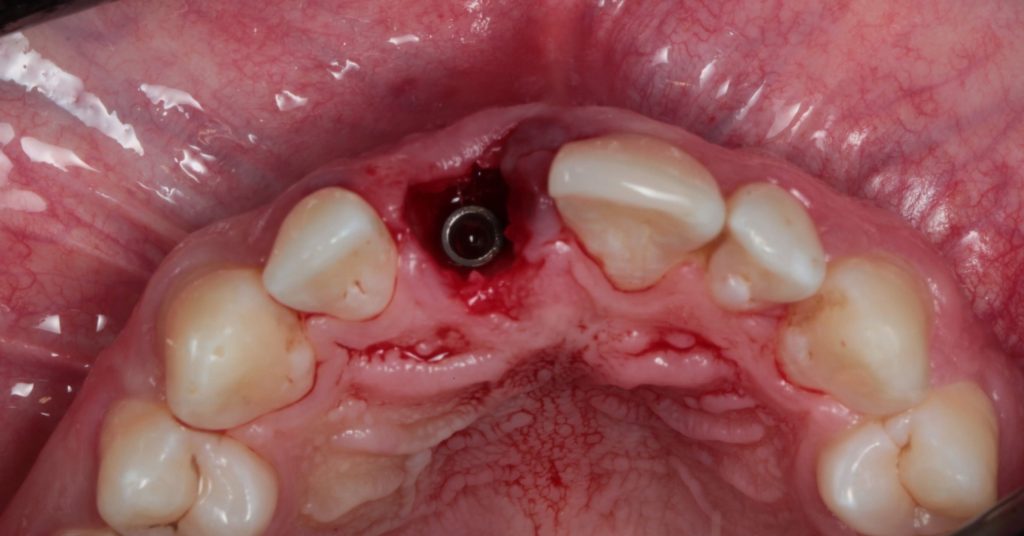
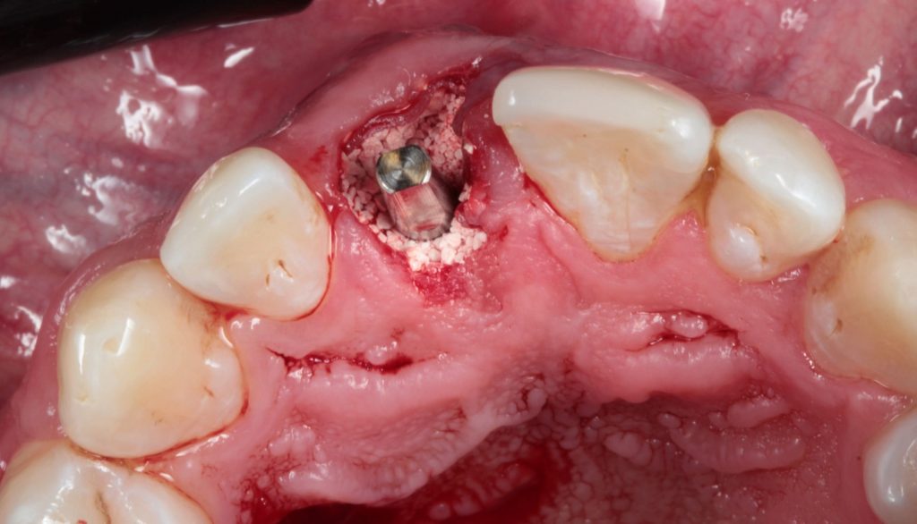
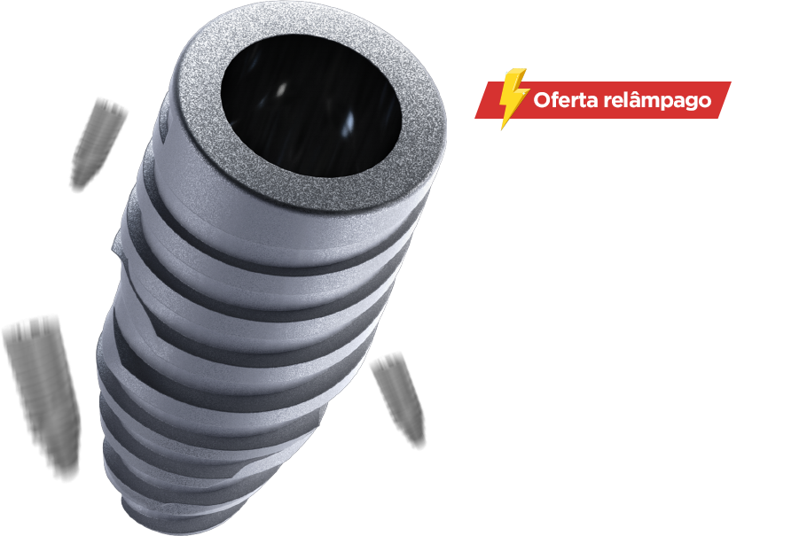
Amacrogeometry of Arcsys implants, with cylindrical body and conical apex as well as trapezoidal threads, favor primary stability, considerably increasing the rates of immediate provisioning. In the present case, a primary stability of 60 N.cm enabled the realization of immediate esthetics (Figure 2).
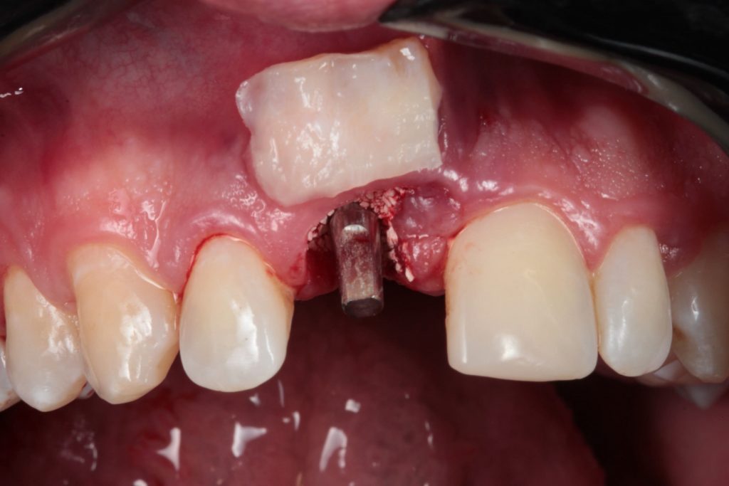
After the installation of the implant, reconstruction of the alveolus was performed with the positioning of a reabsorbable membrane (Genderm, Baumer) and filling of the vestibular gap with 0,5g of Nanosynt FGM synthetic bone substitute (Figure 3).
The presence of a healthy gingival tissue around dental implants, with adequate keratinized tissue band, is considered one of the primary factors not only for esthetics, but mainly for long-term success (Gomes. 2015). In order to improve the quality and esthetics of periodontal soft tissues, a graft of subepithelial connective tissue was performed, removed from the palate by the Zucchelli technique and positioned on the recipient area by tunneling the peri-implant tissues [Figure 4].
In the same session, the prosthetic component for cemented prosthesis was installed (Arcsys FGM 3 x 6 x 3.5 Abutment for Cement-retained Restoration) and a temporary prosthesis was made on Arcsys FGM 3 x 6 multifunctional transfer, where a stock tooth compatible with the size and color of the adjacent teeth was captured with flow resin (Figure 5).
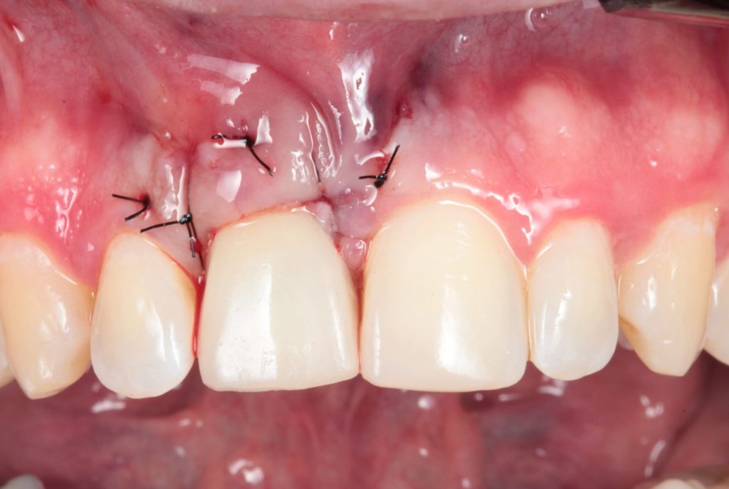
At the end, temporary cementation was performed with Temp Bond and suspensory suture for coronal traction of the flap. After 15 days the patient returned for suture removal and postoperative evaluation (Figure 6).
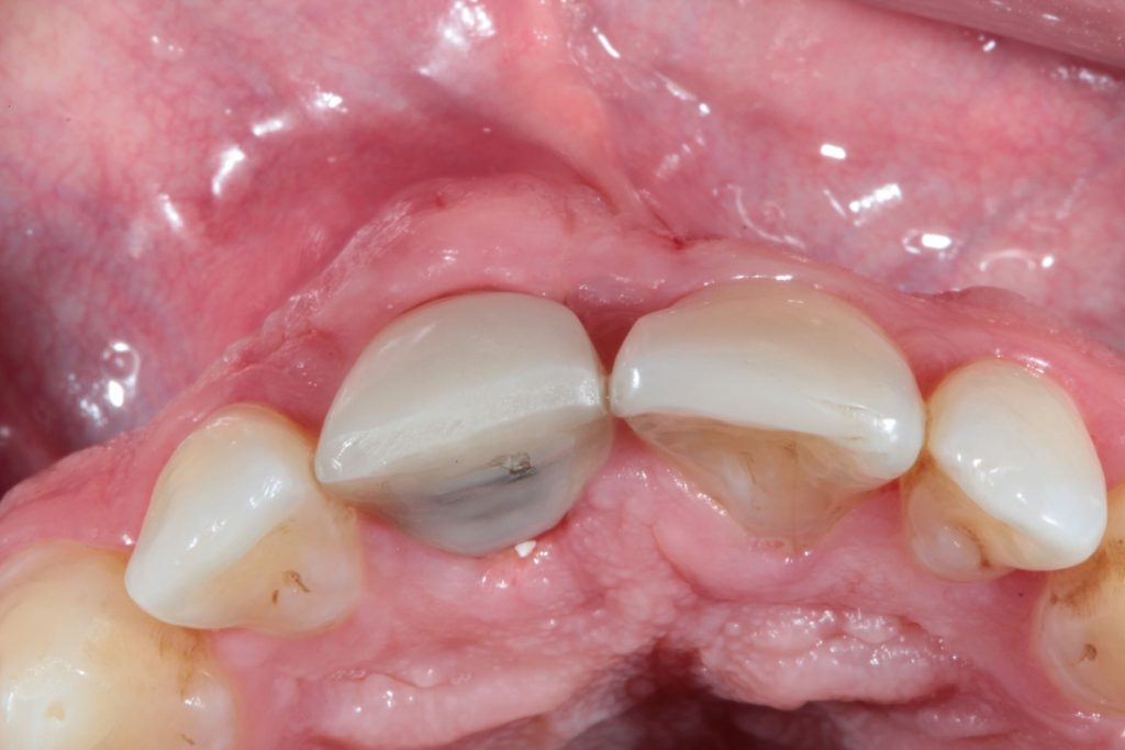
After 90 days of osseointegration and maturation of the periimplant tissues (Photo 7), the patient was discharged and given the go-ahead to receive the definitive crown.


FINAL CONSIDERATIONS
Through this case report, it is concluded that the technique of immediate implantation, associated with reconstruction of the vestibular wall, subepithelial connective graft, and immediate provisioning is safe and effective, decreases the number of consultations and treatment time and maximizes esthetic clinical results of maintenance of the bone and gingival architecture of the peri-implant tissues.

![Implante imediato com provisionalizacao 11 - Immediate implant with immediate provisioning and connective tissue graft: a case report Implante-imediato-com-provisionalizacao-1[1]](https://srv01.fgmdentalgroup.com/wp-content/uploads/2022/11/Implante-imediato-com-provisionalizacao-11.jpg)

