![Imagem1[1]](https://srv01.fgmdentalgroup.com/wp-content/uploads/2022/12/Imagem11.png "Imagem11 - Dento-alveolar rehabilitation with Arcsys implant and Nanosynt bone graft")
Authors: Dr. Jéferson Fagundes
Patient gender and age: Female patient, 67 y.o.
Chief complaint: Absence of tooth 12 and aesthetic dissatisfaction
Initial evaluation: After anamnesis, clinical and tomographic examination, a thin alveolar bone structure was observed in the region of element 12 and volume loss of soft tissue, indicating tissue reconstruction associated with implant installation.
Treatment performed
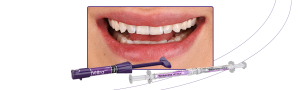
A 3.3×9 Arcsys implant was installed in the region of the element 12, with a torque obtained of 50Ncm. After the implant was installed, a vestibular bone wall fenestration was observed (which was expected, due to the thin bone wall). Soon after, the angular referrer was attached to the implant to personalize the angle of the 3x6x2.5 Arcsys sleeve. After customizing the trunnion to the Arcsys angulator in 6 degrees, the prosthetic component was installed on the implant with the hammer. Then, the autogenous conjunctive graft and the Nanosynt bone graft were applied to the buccal bone wall and protected by an absorbable collagen membrane. The provisional crown was made directly on the multifunctional peek transfer with flow resin. The surgery was completed using PTFE sutures. After 120 days, definitive restorative procedures were initiated, by making ceramic zirconia / feldspathic crowns on teeth and on the implant 12.

FIG. 1 – Initial aspect

FIG. 2 – Initial tomographic section

FIG. 3 – Initial lateral aspect

FIG. 4 – Incisal initial aspect

FIG. 5 – 3,3 x 9 Arcsys implant

FIG. 6 – Implant installed 2mm infra-bone

FIG. 7- Incisal view of the implant

FIG. 8- Angle referrer coupled

FIG. 9- Angulation determined for the abutment

FIG. 10- Angulation customized in 6 degrees

FIG. 11- Abutment installed and Bone graft

FIG. 12- Nanosynt bone graft

FIG. 13- Collagen membrane placed

FIG. 14- Multifunctional transfer over the abutment

FIG. 15- Provisional crown installed

FIG. 16 – Surgery completed

FIG. 17- Lateral postoperative appearance

FIG. 18 – Panoramic image after 30 days

FIG. 19- Clinical aspect after 90 days

FIG. 20- Digital prosthetic planning

FIG. 21- Ceramic crowns on the model

FIG. 22- Cemented crowns: case completed

FIG. 23 – Lateral view: case completed

FIG. 24 – Zoomed image element 12

FIG. 25 – Before/After: lateral view

FIG. 26 – Before/After: frontal view