Authors: Dr. Thiago Roberto Gemeli, Dr. Bárbara Robaskievicz and DP Arnã Ariel da Costa
20-year-old male patient.
QUEIXA PRINCIPAL:
Esthetic compromised by diastemas in the upper arch.
Several anatomical conditions associated with the size or shape of the teeth can generate esthetic disharmonies and negatively interfere with the socialization and selfesteem of many individuals. The absence of contact between adjacent teeth, a condition known as diastema, is commonly observed between antero-superior elements.
The therapeutic approach depends on the diagnosis and can be orthodontic, restorative, or both. Although indirect laminates can also heal this type of condition, the need for dental wear may contradict this option, especially in the case of young patients and healthy elements.
Direct adhesive procedures are an excellent option when well indicated as they are capable of preserving the dental structure and provide excellent esthetic-functional performance. Thus, rehabilitation with composite resins is an important and affordable means to rehabilitate patients with local or generalized diastemas.
CASE REPORT
A 20-year-old male patient attended a private practice complaining mainly of the aesthetic damage associated with the diastemas present in the upper arch. After the anamnesis and initial clinical evaluation, the patient was submitted to complementary photographic/radiographic examinations and archway scanning in order to plan the operative practice.
Because the patient did not want to undergo orthodontic treatment, an exclusively restorative intervention was proposed with an antero-superior six-element approach.
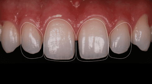
The photographic shots were shared with the laboratory responsible for the digital diagnostic wax-up. The manipulation in software allows editing and sending them to the DP, facilitating communication and allowing them to reach an understanding of what the dentist intends to perform.

Fig. 1 Initial condition of the patient.

Fig. 2 – Initial intra-oral frontal image. |

Fig. 3 Previous elements with reference design for digital wax-up.
After the approval of the project, molding with condensation silicone made it possible to obtain a plaster model. This was scanned and subjected to digital modeling. After final approval, working models were printed and allowed the manufacture of a silicone guide. This facilitates the beginning of the restorative process and the consequent replication of the previously approved anatomical model.


Figs. 4, 5 and 6 – Molding and printed model of the previously approved digital waxing and silicone guide to enable replication of the printed design in the mouth.

Fig. 7 Acid etching for 15s of elements 13 to 23 (Condac 37 FGM).

Fig. 8 Frictional application of adhesive system (Ambar APS FGM).

Fig. 9 Custom silicone guide positioned to assist in the restorative step.
A single resin composite was selected (Vittra APS Unique – FGM) for this work. Its optical properties favor and optimize mimicry with adjacent tissue structures, an extremely favorable feature for daily clinical practice, as it provides practicality, cost reduction, profitability, and versatility of use.
The use of the Ambar APS adhesive seems to be fundamental for the best performance of the composite, as its low camphorquinone load favors mimicry and hardly interferes in the propagation of the color of the substrate to the resinous mass. Another relevant factor to consider is the simplification of the technique, as it dispenses with stratification even in challenging cases.


Figs. 10,11 and 12 Polishing composites with sanding discs (Diamond pro FGM

Fig. 13 Instant shade taking

Fig. 14 Clinical condition after the procedure (A1)

Fig. 15 Shade taking after 10 days of use of the whitening gel.
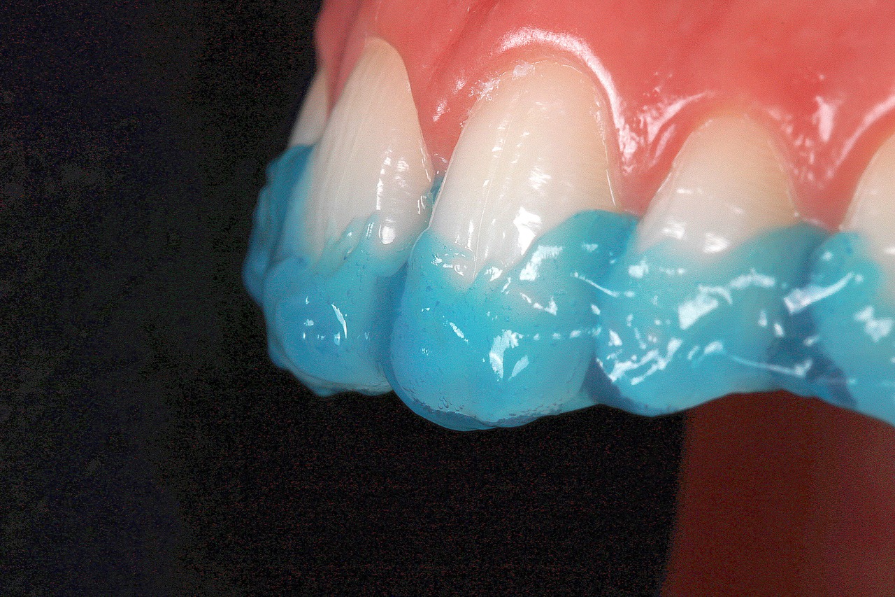
After six months of preservation, the patient showed interest in having his teeth whitened, an option discarded by him in the initial approach. Considering the mimetic ability to propagate the color of adjacent walls, the treatment plan included prophylaxis followed by molding to make the custom molds without having to repeat the plastic intervention. The selected gel (Whiteness Perfect 10% FGM) was recommended for a period of 60 minutes daily for 10 days.
The clinical reassessment showed that the change of color of the substrate by the whitening agent did not impair the final esthetic result of the composite. Unlike what occurs in other resinous systems, the mimetic behavior of the composite used allowed its maintenance
in the mouth, not requiring partial or total repairs after the whitening treatment. This case report is a clear example of how the direct restorative technique with composite resin, when well indicated, is efficient and conservative for closing diastemes, making it an excellent clinical option for the dentist. Another relevant observation is regarding the favorable chameleonic optical behavior of the composite used, which was able to mimic in different optical environments, including when a more significant change in the dentin substrate was promoted (whitening treatment).
REFERENCES
- Santos -Pinto, A. dos; Paulin, R.F.; Martins, L.P. Tratamento de diastema entre incisivos centrais superiores com aparelho fixo combinado a aparelho removível: casos clínicos. J Bras Ortodon Ortop Facial, Cur
- Furuse, A.Y.; Franco, E.J.; Mondelli, J. Esthetic and functional restiration for na anterior relationship wlit multiple diastemata: a multidisciplinary approach. J Prosthet Dent; 99(2): 91-4, 2008.
- De Araujo, E,M.JR.; Baratieri, L.N.; Monteiro, S.JR.; Vieira, L.C.; DE Andrada, M.A. Direct adhesive restoration of anterior teeth: Part2. Clinical
- protocol. Pract Proced Aesthet Dent. Jun; 15(5): 351-7; quis 9, 2003.

![Plastias anteriores em resina composta com posterior alteracao do substrato dentario1 - Anterior rehabilitation in composite resin with subsequent change of the dental substrate Plastias-anteriores-em-resina-composta-com-posterior-alteracao-do-substrato-dentario[1]](https://fgmdentalgroup.com/wp-content/uploads/2022/11/Plastias-anteriores-em-resina-composta-com-posterior-alteracao-do-substrato-dentario1.jpg)


