Raphael Vieira Monte Alto, Marco Antônio Gallito e Gustavo Oliveira dos Santos
INTRODUCTION
Cementation is a fundamental step in the clinical protocol of indirect restorations and has been increasingly refined with the emergence of new cementing agents. An ideal cementation material should possess satisfactory mechanical properties, adhesion to dental structures and restorative materials, low solubility, appropriate viscosity, among other characteristics.
Initially, ceramics were cemented using zinc phosphate cement or glass ionomer cements. Such cements often led to the failure of these restorations due to displacement, marginal leakage, or aesthetic issues. With the advent of composite resins and adhesive systems, the development of resin cements became feasible. These cements have the same composition as composite resins, consisting primarily of an organic matrix and filler particles treated with silane. They exhibit adhesive properties, the necessary flow for cementation, mechanical strength, aesthetic properties, and are insoluble in the oral environment. For these reasons, resin cements are currently the most commonly used materials for ceramic restoration cementation. In addition to these characteristics, resin cements exhibit adhesion to dental structures when used in conjunction with adhesive systems, and to indirect aesthetic restorative materials when they are silanized.
Resin cements can be self-activated, light-activated, or dual-cured. Self-activated cements are used for cementing all types of prostheses. Light-activated cements are mainly employed for cementing ceramic veneers, while dual-cured cements are preferred for cementing ceramic restorations and prefabricated posts due to their slow chemical activation and extended working time. When exposed to light, these cements achieve rapid setting, thus promoting greater conversion of monomers into polymers. The aim of this study was to report the clinical sequence of adhesive cementation of a pure ceramic aesthetic restoration using the dual-cure resin cement AllCem (FGM).
CEMENTATION SEQUENCE
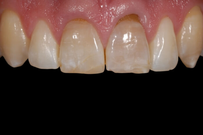
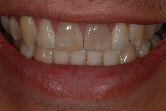
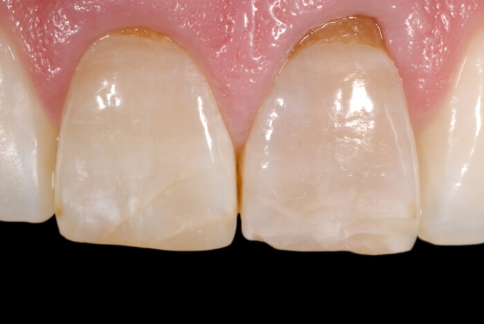
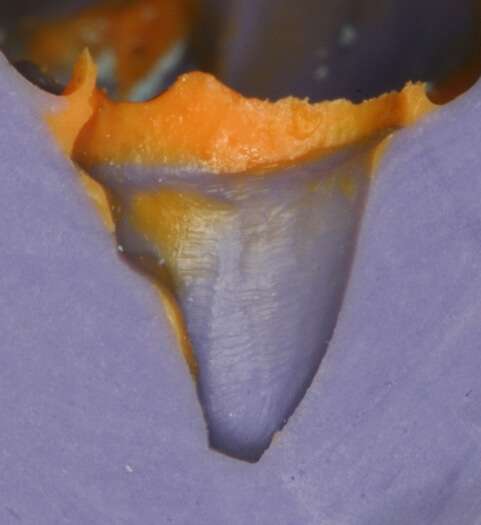
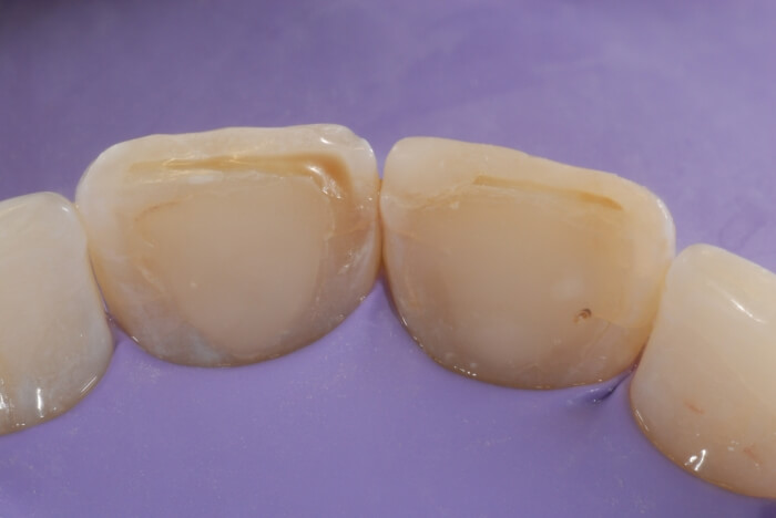
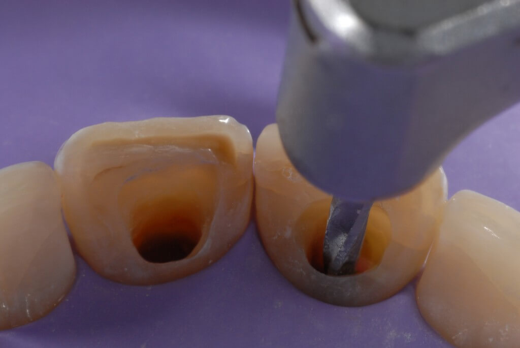
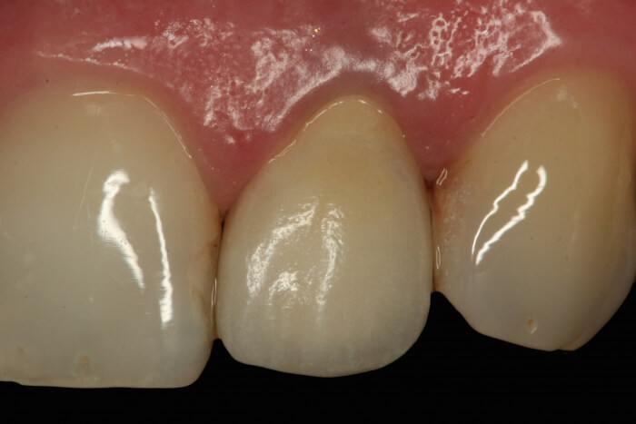
The patient was dissatisfied with their smile due to a full acrylic resin crown with color alteration on tooth 22 (Fig. 1). Initially, the removal of this restoration was performed (Fig. 2), followed by refinement of the preparation for the fabrication of a metal-free ceramic full crown (Fig. 3). Subsequently, an impression was taken using addition silicone due to its high dimensional stability (Fig. 4), and immediately after, a provisional restoration was fabricated using bis-acrylic resin (Fig. 5). The shade selection was carried out using the Linear Guide 3D Master shade guide (Vita, Germany), and the impression was immersed in 2% glutaraldehyde for 10 minutes for disinfection before being sent to the prosthetic laboratory. There, the model was obtained, the coping was fabricated, and the veneering ceramic was applied (Figures 6 and 7).
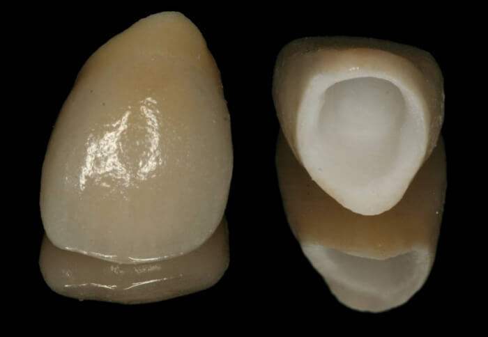
The restoration was placed in the mouth, and its proximal surfaces were adjusted, while occlusal movements were replicated to identify potential premature contacts and interferences. After a final assessment of color, shape, and texture, the work was deemed suitable for cementation.
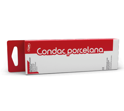
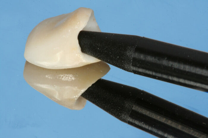
Consequently, the prosthetic piece was cemented. Internal conditioning of the piece was performed using a porcelain conditioner based on 10% hydrofluoric acid (Condac Porcelana, FGM, Brazil) for 20 seconds (Fig. 8 and 9). After this step, the piece was thoroughly washed to completely remove the product and dried with air jets, presenting an opaque appearance, characteristic of the correct conditioning pattern (Fig. 10).
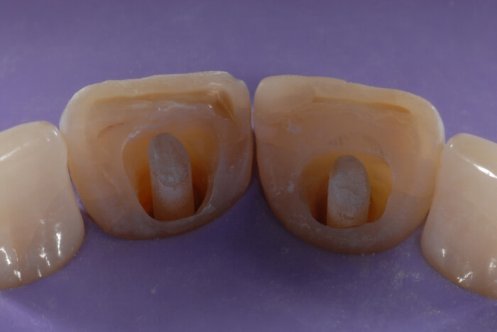
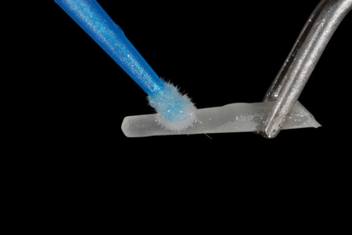
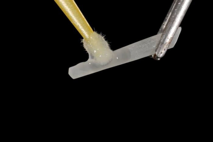
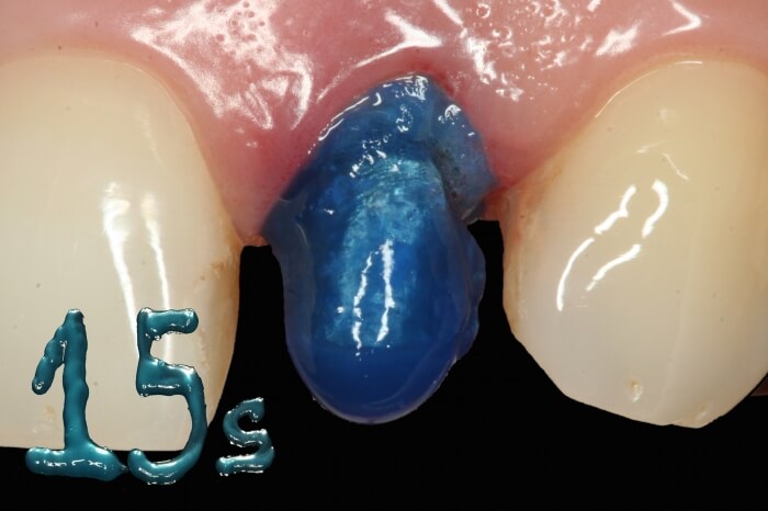
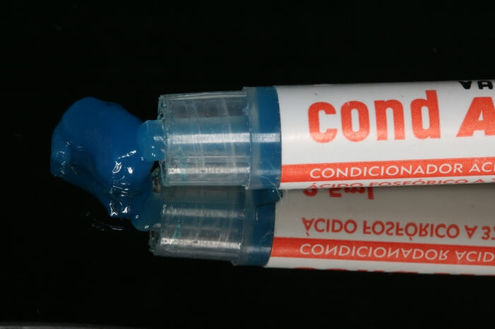
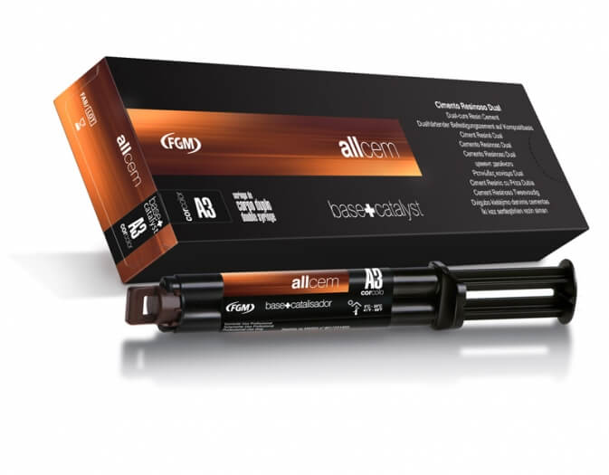
Next, the silane agent Prosil (FGM) was applied with the aid of a Cavibrush tip (Figures 11 and 12). After silanization was completed, the surface of the piece appeared shiny and was ready for definitive cementation. Conditioning of the residual tooth structure was achieved using 37% phosphoric acid Condac 37 (FGM, Brazil) for 15 s (Figures 13 and 14). Following conditioning, the surface was washed, dried, and an adhesive system was applied to the residual tooth structure. The cement color (A3) was chosen and it was placed inside the piece using an automixing tip (Figures 15 and 16), and the ceramic crown was guided into place. Excess material was removed, and polymerization was performed using a photopolymerization device (Figure 17).
DISCUSSION
The adhesive cementation technique demands the operator’s knowledge about the adhesion mechanism. To achieve success in the procedure, it is necessary to treat the remaining dental structure and the internal surface of the indirect restoration prior to cementation.
Conditioning of the remaining dental structure with 35 to 37% phosphoric acid provides demineralization in both enamel and dentin, aiming for hybridization. In enamel, there is dissolution of the center or periphery of the prism head, and in dentin, partial demineralization of the peritubular and intertubular dentin occurs, exposing collagen fibers and thus promoting the formation of the hybrid layer, responsible for adhesion.
Ceramic conditioning promotes the chemical-mechanical bonding between the dental structure and the internal surface of the prosthetic piece. 10% hydrofluoric acid (Condac Porcelana) is used for this purpose and acts on the vitreous phase of some ceramics. This conditioning is important for an interaction between the resin cement and the ceramic restoration, thus increasing retention and restoration strength.
Silanization is performed after conditioning the internal surface of the restoration and is of fundamental importance to chemically match this surface with the resin cement. Prosil (FGM) is an ethanolic solution of 3-Methacryloxipropyltrimethoxysilane hydrolyzed, which can be used as a bonding agent in adhesion and cementation processes of ceramic pieces, ceromers, and glass fiber posts, thereby increasing adhesive capacity by up to 30%. This silane promotes the bond between interfaces due to its high reactivity with methacrylic monomers and surfaces containing inorganic elements.
The resin cement AllCem (FGM) is a dual-curing cement, radiopaque, and with excellent aesthetic properties. The combination of both curing mechanisms, light-activated and chemically activated, ensures the polymerization of the product in situations with and without light access. This cement also presents excellent adhesive and mechanical properties, as well as ease of application due to the dual-barrel syringe. The use of this syringe ensures the correct proportioning of the product, and the automix tips ensure the homogeneity of the pastes, preventing the incorporation of bubbles, which greatly contributes to the effectiveness of cementation. In addition to these characteristics, it is important to emphasize that care must be taken during excess removal.
FINAL CONSIDERATIONS
Adhesive cementation is a crucial step in achieving success with indirect restorations. The adhesive cementation products Condac Porcelana (FGM), Prosil (FGM), and AllCem (FGM) demonstrated their effectiveness in the cementation of the mentioned indirect restoration in the presented clinical case.
REFERENCES
- Agra, C.M.; Vieira, G.F. Quantitative analysis of dental porcelain surfaces following different treatments: correlation between parameters by a surface profiling intrument. Dent. Mater J. v.21, p.44-52, 2002.
- Cura, C.; Matsumura, H.; Atsuta, M. Effect of different bonding agents on shear Bond strengths of composite-bond porcelain to enamel. J. Prosthet. Dent. v.89, n.4, p.394-399, 2003.
- Chen, J.H.; Matsumura, H.; Atsuta, M. Effect of different etching periods on the bond strenght of a composite resin to a machinable porcelain. J. Dent. Res. v.28, n.1, p. 53-58, 1998.
- Chen, J.H.; Matsumura, H.; Atsuta, M. Effect of etchant, etching periods and silane priming on bond strength to porcelain of composite resin. Oper Dent. v.23, n.5, 1998.
- Sheet, J.J.; Jensen, M.E.: Cutting interfaces and materials or etched porcelain restorations. A status report or the American Journal of Dentistry. Am. J. Dent. v.1, n.5, p.225-235, 1988.
- Badini, S.R.G. et al. Cimentação adesiva – Revisão de literature. Revistas Odonto. v.16, n.32, p.105-115, 2008.


