In this clinical case, composites resins were used to stratify and simplify the transformation of this patient’s smile.
Author: Dr. Carlos E. Francci, Dr. Alexander C. Nishida and/y Dr. Guilherme de S. F. Anzaloni Saavedra.
27-year-old male patient.
Dissatisfaction with the color and the spaces between the anterior superior teeth.
Initial evaluation
The case offers an opportunity to perform whitening on a yellowish substrate and the re-anatomization of the anterior superior teeth with the new composite Vittra APS Unique.
Treatment performed with composites
The detailed examination allowed for the visualization of an asymmetry between the size of the central and lateral incisors, with an accentuated wear in the canines. The initial shade taking showed that the substrates were very yellowed, indicating the need for whitening for esthetic improvement and patient’s satisfaction. Prophylaxis of the teeth was carried out followed by shade taking with the Vita 3D Master guide.
After molding with addition silicone for the upper arch and alginate for the lower arch, 1mm trays were made of vacuum-forming EVA . After the fitting of the trays, two syringes of Whiteness Perfect 10%, the trays and a case for the trays were handed to the patient. The upper model in special type 3 plaster, obtained with the high pressure of the addition silicone was scanned (Intraoral CS 3600 Color Scanner – Carestream) and the digital plan of the smile was done with Exocad ChairsideCAD (Exocad).
The patient was instructed on the use of the gel and to wear it for 2 hours a day. One week after the end of the whitening, the restorative treatment began.
The treatment was conducted under absolute isolation and the restorations were carried out with the new Vittra APS Unique composite to harmonize the smile. The diastema between the incisors was closed, the mesial/distal and incisive/cervical proportions were corrected. A slight rotation of the left upper lateral incisive was also corrected and the canine guidance was reestablished.
Step by step

Fig. 1 – Photograph of the smile revealing the relations between the face and the teeth, initial step in search of horizontal (bi-pupillary in this case) and vertical (sagittal median line) references.

Fig. 2 – Shade taking of the central incisors using the Vita 3D Master guide. The shade taken was 1M2.

Fig. 3 – Shade taking for upper canines using the Vita 3D Master guide. The shade taken was 2L2.

Fig. 4a – The shade taking drew the attention to a great amount of scratches on the vestibular enamel, especially on the incisors, due to the inadequate removal, with a diamond drill, of the resin cement from the fixation of brackets.

Fig. 4b – Use of silver powder to better evidence the scratches made by the diamond drills on the surface of the enamel of the anterior teeth.

Fig. 5 – To decrease the amount of scratches and remove the remaining resin cement from the fixation of the brackets, a 22 blade multi-laminated drill was used with countersink multi-applicator in 8000 rpm.

Fig. 6 – The patient’s teeth were molded and a set of upper and lower trays was made of EVA for the at-home whitening treatment.

Fig. 7 – The patient was given three syringes of Whiteness Perfect 10% (carbamide peroxide at 10%) for the at-home whitening treatment with the use of the trays.

Fig. 8 – After 3 weeks, the evolution of the whitening treatment showed very satisfactory results with the incisors reaching shade 0.5M1, the second lightest in the Vita Bleached Guide.

Fig. 9a – During whitening, the models were scanned (intraoral CS 3600 Color Scanner – Carestream) and the digital plan of the smile was carried out in the upper arch. Note that a semi-transparent overlapping of the final smile was done to visualize what was really needed in terms of re-anatomization with composite.

Fig. 9b – In this screen of Exocad, the same plan was done with greater opacity for the modification of the composite, showing where the enamel would be apparent, without the cover of the composite.

Fig. 10 – For the beginning of the restorative procedure, absolute isolation was carried out with the use of a rubber dam for controlling moisture and to improve the vision of the operation site.

Fig. 11 – Enamel etching using Condac 37 (phosphoric acid at 37% – FGM) on the whole vestibular surface of the upper right incisor in order to guarantee the adhesion of the composite to any part of that tooth. The protection of the adjacent teeth was mace with teflon tape.

Fig. 12 – After abundantly washing and drying of the enamel, the Ambar Universal APS adhesive was applied with the help of a long Cavibrush, which better spreads the adhesive on the wide vestibular surface. It is possible to see that the adhesive is almost transparent for having low levels of camphorquinone.

Fig. 13 – Photopolymerization is potentialized by the presence of APS (Advanced Polymerization System) in the formula of the Ambar Universal APS adhesive.

Fig. 14 – An increment of Vittra APS Unique composite was taken to the vestibular surface and sculpted gently.

Fig. 15 – The handling of the composite is easier for its low stickiness.

Fig. 16 – The composite was sculpted with the help of a spatula with a non-adhering rolling tip which rolls to spread the mass in a uniform way, without air bubbles.

Fig. 17 – The aspect of the central incisor with its finalized primary anatomy. The Vittra APS Unique composite mimicked the color of the substrate without showing any difference between the enamel of the tooth and the restoration in composite, even without beveling.

Fig. 18 – The same process was repeated for the construction of tooth 21. In the case of central incisors, it is essential that the teeth are spectacular images of one another in order to provide harmony.

Fig. 19 – After restorations were made in the lateral incisors for the closing of the remaining spaces, repeating the same protocol for the preparation, adhesion and construction, the strategic additions were made in the incisal surfaces of the canines to redefine the canine guidance and to harmonize the incisal interdental Spaces.

Fig. 20 – Small corrections were made and excess material was removed with the use of Diamond Pro disks according to the sequence starting with the most abrasive (darker blue) to the least abrasive (lighter blue or white).


Figs. 21a and 21b – Pre-polishing was carried out with pastes Diamond ACI and ACII using the Diamond felt disk.

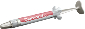
Fig. 22 – The Diamond R (FGM) (12µm) paste was used for polishing with humid felt disks Diamond and Diamond Flex.

Fig. 24 – Smile at the end of the treatment. The whitening treatment illuminated the substrate and the closing of the spaces allowed for harmony in the whole smile.


Figs. 25a and 25b – Details of the reanatomization with the composite Vittra APS Unique seen from the right and from the left.
El verdadero efecto camaleónico de Vittra APS Unique


